Zoologists have found that spiny mice and representatives of three other closely related genera of rodents have osteoderms—cutaneous ossifications—in the skin of their tails. Such structures are found in many reptiles, such as crocodiles and turtles, but among modern mammals they were until now known only from armadillos. 
Apparently, osteoderms serve rodents for protection. If a predator grabs a spiny mouse by the tail, it will shed its skin like a stocking and run away. Ossification in the skin makes this strategy more effective, ensuring that the enemy cannot sink its teeth into deeper, non-flaking tissue. The results of the study were published in an article for the journal iScience.
Osteoderms are called ossifications located in the skin of some vertebrates. As a rule, these are small, flattened formations that perform a protective function. In some cases, they even grow together, forming a full-fledged shell. It is assumed that osteoderms arose independently in different evolutionary lineages of amniotes at least nineteen times. They are especially common in reptiles. For example, osteoderms underlie the horny scutes of crocodiles and turtles and the scales of some lizards. However, birds do not have such structures, and among living mammals only armadillos (Cingulata) have them, in which osteoderms form the shell (some authors also classify the bone base of the horns of artiodactyls as osteoderms).
A team of zoologists led by Malcolm Maden from the University of Florida has described another example of osteoderms in modern mammals. The researchers focused on rodents from the subfamily Deomyinae: spiny mice (Acomys), wire-haired mice (Lophuromys), Congo mice (Deomys) and large-toothed mice (Uranomys). They are common in Africa, the Middle East and the Mediterranean islands. 
After studying museum specimens of spiny mice and their relatives using computed tomography, Maden and his co-authors discovered unusual structures in their tails - overlapping ossifications located in the dermis, shaped like tiles. They cover the entire tail, forming rows of rings surrounding the vertebrae and spinal cord, each of which has eight to eleven separate ossifications. Ossifications located close to the body are as dense as bones, while those located closer to the end of the tail are much less dense. In shape and structure, these structures are very similar to osteoderms on the backs of armadillos and on the tails of Elgaria hosmeri lizards. Having compared all the information obtained, the authors came to the conclusion that the ossifications located in the tails of rodents from the subfamily Deomyinae can be considered osteoderms.
The osteoderms of adult spiny mice have lacunae containing osteocytes. In addition, neurovascular channels with capillaries in them pass through the ossifications. The dorsal surface of each osteoderm is adjacent to the epidermis, from which it is separated by a layer of dermis three to four cells thick. And the ventral surface of the ossifications is immersed in connective tissue or in contact with adipose tissue. In this case, hair follicles are located between the osteoderms, and three hairs emerge from under each plate. 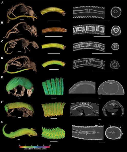
At the next stage, Maden and his colleagues scanned several spiny mice of different ages and studied sections of their tails. It turned out that in newborn rodents osteoderms are present only at the base and in the middle part of the tail. Only at the age of six weeks, when approaching puberty, ossifications appear at the end of the tail, first in the form of thin plates.
Each ring of osteoderms begins to form from the dorsal part of the tail. The first ossifications are laid here - and then they spread to the ventral side. In this case, the formation of each osteoderm begins with several fibroblast-like cells lying under the epidermis, which differentiate into osteoblasts. The individual osteoblasts then assemble and synthesize the bone matrix, eventually creating a flat lamina with osteocytes inside. Osteoderms of armadillos are formed in a similar way.
To determine the molecular mechanisms behind osteoderm formation, the researchers performed transcriptomic analysis of samples taken from the proximal, middle, and distal tails of newborn spiny mice. It turned out that in the mouse tail, gradually, from base to tip, there is a transition from hair formation to bone development. Among the twenty genes whose activity is most suppressed in the proximal part of the tail, sixteen are responsible for hair growth and keratin production. And among the twenty genes whose expression is especially increased in this part of the tail, twelve are associated with bone growth. Additional analysis of signaling pathways showed that osteoblast differentiation, connective tissue differentiation, and bone mineralization are actively occurring in the proximal part of the tail.
Why spiny mice and their relatives need osteoderms in their tail is not yet fully understood. The authors suggest that this may be due to their defense mechanism. If you grab a spiny mouse by the tail, the skin peels off like a stocking. This skin will remain in the predator's mouth - and the rodent will be able to escape. Osteoderms may make this strategy more effective by ensuring that the predator cannot pierce the skin and sink its teeth into deeper tissue that is no longer peeled off. A similar function is performed by ossifications in the skin of Madagascar large-scaled geckos (Geckolepis).
Maden and his co-authors note that until now, studying the cellular and molecular mechanisms that are responsible for the formation of osteoderms has been difficult. The fact is that previously known animals with such ossifications are not very well suited for captivity and reproduce rather slowly. However, spiny mice do not have these disadvantages, so they can become an ideal laboratory model for studying osteoderms.
One of the reptile species with well-developed osteoderms is the Komodo dragon (Varanus komodoensis). In adult individuals of this species, the bony plates in the scalp form real chain mail, which serves as protection from relatives.
0 comments